You will find below a short description of the scientific expertise within the Aurastem network.
UNDER CONSTRUCTION! This list is not exhaustive. To be referenced on this website, please complete this form
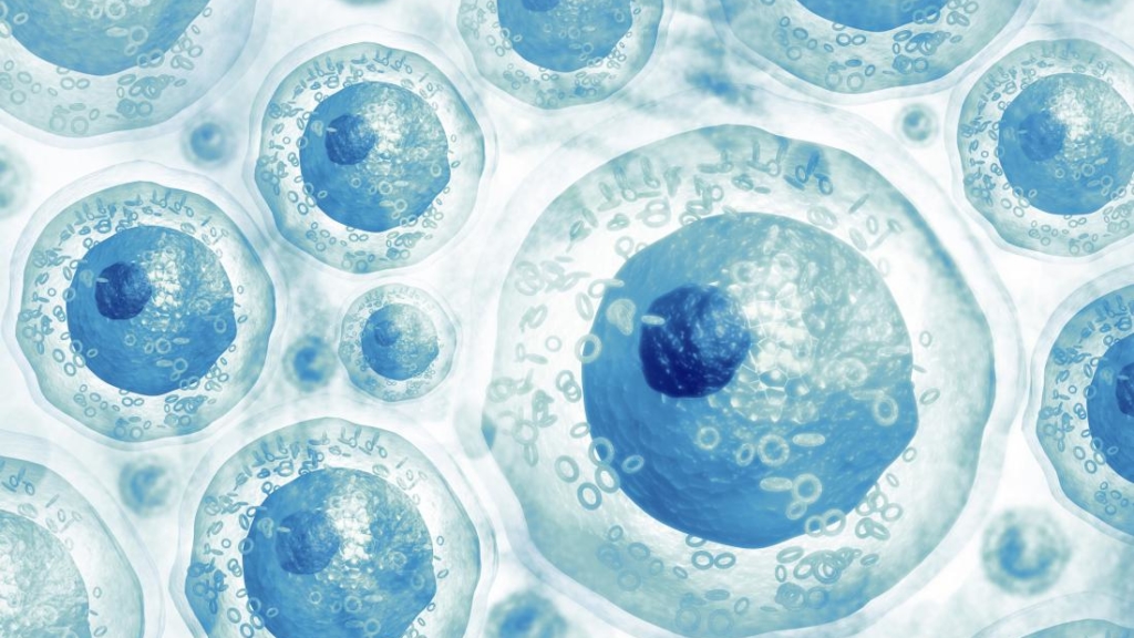
Pluripotent stem cells
Somatic stem cells

PLURIPOTENT STEM CELLS
CLAIRE CHAZAUD – GReD Institute, Clermont-Ferrand
Team « Molecular mechanisms of cell lineage differentiation in the early mouse embryo »
Our team is interested in deciphering the molecular mechanisms regulating the first cell differentiation event that takes place in the early mouse embryo. We use mouse embryos as well as stem cells models (ES cells, F9 cells).
Methodologies: embryo cultures, immunoFISH, single cell RTqPCR, proximity ligaion assays.
Biological resources: transgenic mice (Gata6tm2.1Sad, Nanogtm1Yam, Sox17CreERT2, Zp3-Cre, Igf1rtm1Jcbr, Insrtm1Khn); cell lines: Gata6-/- Es cells.
FABRICE LAVIAL – Cancer Research Center, Lyon
Team « Cellular reprogramming and oncogenesis »
The lab is interested in deciphering the molecular mechanisms that control cellular identity in various steady-state and pathological contexts, ranging from mouse peri-implantation development to reprogramming and tumorigenesis.
Methodologies: iPS/ES cell cultures, 3D organoid cultures, omics approaches (CIP-seq, RNA-seq, ATAC-seq), bioinformatics.
Models: embryos and mice.
CHRISTOPHE MARQUETTE – Molecular and Supramolecular Chemistry & Biochemistry Institute (ICBMS)
Team « 3D.FAB »

The platform aims at implementing its 3D bioprinting know-how to stem cell research. We generate proliferative 3D structured material for both tissue generation and stem cell production.
Methodologies: 3D printing of bone-marrow and iPS thanks to in-house developed bioikns and bioprinting methodologies.
Biological resources: 3D FAB printing platform, cytometry, cell culture infrastructures.
DAVID VOLLE – GReD Institute, Clermont-Ferrand
Team « Environment, Metabolism, spermatogenesis, pathophysiology and inheritance »
Our aim is to elucidate the cellular and molecular mechanisms that support the harmful effects of altered metabolism and/or exposure to environmental molecules on male reproductive functions.
Methodologies: We achieve these goals using a combination of high-throughput technologies (RNAseq, ChIP-seq, metabolomic) to investigate differential metabolic pathways and gene expression profiles from model cell lines, genetically modified animals (mouse, nematode). Histological approaches, FACs cell sorting with single cell analysis technologies, cell culture and imaging as well as bio-informatics.
Models: Rodent models for endo- and xenobiotics (TGR5F/F; FXRF/F; CAR-/-; PXR-/-); Rodent models for cancer studies: PtenF/F; KRASG12D; Human and mouse immortalized germ cell lines; Nematodes and and access to siRNAbiobank

PIERRE SAVATIER – Stem Cell and Brain Research Institute, INSERM U1208
Team « Pluripotent Stem Cells in Mammals »
Developing the technology of embryo chimeras with pluripotent stem cells for developmental studies in primates.. Molecular mechanisms underpinning naive pluripotency and chimeric competence in rabbits and primates.
Methodologies: In vitro fertilization in non-human primates ; Culture and genome editing of pluripotent stem cells ; embryo chimeras ; bi-photon live imaging ; RNA-sequencing
Models: Macaque monkey and rabbit facilities ; embryo-derived and induced pluripotent stem cells in rabbits, marmosets, rhesus, chimpanzees and Humans.
CHARLENE GUILLOT – GReD Institute, Clermont-Ferrand
Team « Dynamic Regulation of Body Axis Morphogenesis »
Our primary interest is to understand the role of Environmental Cues in Axis Morphogenesis via studying the Axis Stem Cells (Neuro Mesodermal Progenitors)
Methodologies: histology, fate mapping approaches (single cell color barcoding), transcriptomic approaches including single-cell RNA-seq, In-ovo live imaging.
Resources: Chicken and Quail eggs. Leica Thunder Model Organism.
charlene.guillot(at)uca.fr ou charlene.guillot(at)inserm.fr
SOMATIC STEM CELLS
OLIVIER RAINETEAU – Stem Cells and Brain Research Institute (SBRI), Lyon
Team « Development & plasticity of the postnatal forebrain »
Our main interest is to understand the potential (but also limitations) for postnatal neural stem cells to participate to tissue repair.
Methodologies: histology, fate mapping approaches (BrdU, electroporation, transgenesis, …), transcriptomic approaches including single-cell RNA-seq.
Biological resources: transcriptional datasets of postnatal forebrain NSCs, transgenic mice for NSCs visualization and/or manipulation.
LYDIA CAMPOS – University Jean Monnet & University Hospital
Our research follows 2 different axes: 1) Mechanisms of Leukemogenesis in the context of acute myeloid leukemia (AML). More specifically we are interested in the role of Embryonic Stem Cell markers, including the factors OCT4, SOX2 and NANOG in the etiology of AML, especially at the level of the Leukemic Stem Cell and their prognostic capacity for the progression of the disease. 2) The role of the Medullary Microenvironment in the pathogenesis of Myelodysplastic syndromes; we are interested in the impact of FAK-deficient stroma on hematopoiesis through direct interactions between these two cells and indirectly through disruption of cellular metabolic activity which can lead to abnormalities in proliferation, differentiation, autophagy, etc:.
Methodologies: Methodologies used Cell cultures, flow cytometry, PCR, NGS, FISH, Karyotyping, cell sorting by magnetic beads
Biological Resources: Tumor Cell bank, leukemic cell lines
EMILIE BRASSET – GReD UMR6293 – UCA – INSERM U1103
Team « INSTABILITÉS GÉNÉTIQUES ET CONTRÔLE PAR LE GÉNOME HÔTE »
Using Drosophila as a model our project aims to uncover underlying specificities of the piRNA pathway in the control of Transposable Elements, in the female gonadic tissue and in some extend in the somatic and germline stem cells, allowing an appropriate balance between genome protection and genome plasticity.
Methodologies: FACS cells sorting, smFISH, immunostaining, ChIP, small RNA sequencing (sRNAPipe), mRNA sequencing, 4C, genetic approaches, CRISPR…
Biological resources: Drosophila melanogaster tissues (more precisely gonads), ovarian somatic cells line.
DAMIEN FREYSSENET – Laboratoire Interuniversitaire de Biologie de la Motricité
Team « Deconditioning Reconditioing of skeletal muscle- Systemic Environment »
The main objective of our team is to understand the mechanisms of skeletal muscle deconditioning and to develop innovative and effective strategies to combat skeletal muscle deconditioning. Intrinsic (inside muscle fiber) and systemic regulatory mechanisms are explored to meet the following 4 objectives: i) to identify the cellular and molecular mechanisms at the origin of skeletal muscle deconditioning, ii) to determine how the systemic environment contributes to skeletal muscle deconditioning, iii) to determine how skeletal muscle deconditioning may contribute to the evolution of the pathological process, and iv) to develop strategies to prevent or limit the extent of muscle deconditioning.
Methodologies: Western blot – Spectroscopic and fluorescence enzymology – RT-qPCR – PCR – Microscopy – Cell culture – Animal physiology and electrophysiology
Biological Resources: Transblot (BioRad) – ChemiDoc MP Imaging system (BioRad) – Skeletal muscle physiology (Aurora Scientific) – CFX96 touch real time PCR detection system (BioRad) – Axio Imager 2 microscope (Zeiss) – Gene electrotransfer GET42 (EIP) – Lecteur microplaque Hidex Sense (LabLogic SciencTec) – Cell culture room
LUCAS WALTZER – Institut de Génétique Reproduction et Développement
Team « Epigenetic regulation and Hematopoiesis »
Our goal is to shed new lights on the fundamental principles that govern gene expression and underlie progenitor differentiation or maintenance. Our projects aim at deciphering the mechanisms that regulate bood cell development and how these mechanisms go awry in patholocial situations. Using Drosophila as a model organism, we are particularly interested in unravelling the function and mode of action of epigenetic enzymes in hematopoiesis and beyond.
Methodologies: Drosophila genetics, imaging, molecular biology (NGS approaches, including RNA-seq, ChIP-seq, SMRT-seq and HiC).
Biological Resources: Transblot (BioRad) – ChemiDoc MP Imaging system (BioRad) – Skeletal muscle physiology (Aurora Scientific) – CFX96 touch real time PCR detection system (BioRad) – Axio Imager 2 microscope (Zeiss) – Gene electrotransfer GET42 (EIP) – Lecteur microplaque Hidex Sense (LabLogic SciencTec) – Cell culture room
ANNE MEY – U1060 INSERM-INRAE 1397
Team « CarMeN Laboratory – Team 1 »
Our works address the question of mesenchymal stem cells as initial targets of overnutrition in the emergence of metabolic diseases..
Research themes: Functional exploration of adipose tissue stem cells (= mesenchymal stem cells) in obesity and metabolic diseases – Influence of obesogenic diet on the functions of adipose stem cells all the life long – Metabolic programming – Perinatal imprinting- Immunometabolism – Influence of the depot of origin on the functions of adipose stem cells…
Methodologies: Isolation and culture of primary stem cells from mouse and human adipose tissues – Diet-induced obesity and metabolic diseases – Comparison of stem cells from subcutaneous and visceral adipose depots – Metabolic phenotyping of animal models – Cell metabolism analysis – Flow cytometry – Co-culture experiments – RNA seq…
Biological resources: Adipose stem cells from mice and humans. Mouse models of obesity.
FLORENCE RUGGIERO – IGFL (UMR5242)
Team « Matrix Biology and Pathology »
We aim (1) at characterizing the ECM environment (composition, topology and physical properties) of stem cells or progenitors in different biological and pathological contexts, (2) at identifying cell-specific gene expression signatures during these processes and (3) at understanding the relationships and functions of various ECM partners in the regulation of cell behavior during organogenesis and tissue regeneration as well as their disturbance in diseases.
Research themes: Biology and Pathology of extracellular matrix (specifically collagens) in development, regeneration and tissue homeostasis. Stem cells related themes: skin and muscle-nerve development and regeneration, ECM environment of Muscle Stem Cell niche (nature and function) in collaboration with P. Mourikis (Paris).
Methodologies: In vitro models (3D Cell culture) and in vivo (zebrafish), living imaging; microcopies; biochemistry, transcriptomic analysis, genome editing
Biological Resources: Isolated cells from mice and zebrafish, developing zebrafish, Targeted tissues: skin and the neuro-muscular system
JEROME LAMARTINE – LBTI – CNRS UMR5305
Team « SKin Functional Integrity (SKIN) »
Our work aims at identifying skin cells sub-population (keratinocytes, fibroblasts, adipocytes) with high potential in tissue regeneration. Addressing the Role of ncRNAs in the maintenance of the epidermal progenitor pool Ambition: to implement single-cell genomics and functional imaging approaches to better characterize the function and dynamics of cellular sub-populations..
Research themes: How the functionality of the healthy skin is maintained? How the skin functionality can be fragilized by environmental changes (diabetes, inflammation, aging)? How the functionality of the compromised skin can be recovered after wound healing?
Methodologies: Skin functional exploration in vivo and in vitro. Lineage tracing in mice, imaging sub-populations in the tissue by IF and sm-RNAFISH. Functional tracing of microRNAs in 3D-organotypic tissues..
Biological resources: Bank of primary cells from human skin. Mouse models of fragilized skin (defects in thermoregulation, defects in skin barrier, inflammation, diabetes)..
Veronique Maguer-Satta – CRCL-UMR 5286-Inserm 1052
Team « BMP, Ecosystem, Stemness & Dynamic in Cancer »
Our goal is to improve our understanding of the mechanisms underlying cancer initiation by revealing how the microenvironment promotes stem cell transformation, expansion of cancer stem cells as well as resistance through adaptation of the tumor ecosystem to treatment pressure and by using the BMP pathway.
Research themes: Normal and tumoral cancer stem cells interactions with their niche.
Methodologies: Breast: mammospheres, CFC, TDLUs assays. Hematopoiesis: single-cell phenotype, dormancy, cell-confinment.
Biological Resources: Primary MSC, HSC and LSC from myeloid leukemias. Primary mammary tissue from normal and breast cancer, Relevant cell lines models modelizing tumor initiation and progression.
ERIKA COSSET – CRCL, UMR U1052 – CNRS 5286
Team « Genetic instabilities and control by the host genome »

Our team focuses on the cellular and molecular mechanisms underlying cancer cell and cancer stem cell addiction to different metabolic pathways, and to design new strategies to exploit these vulnerabilities to halt brain tumour progression..
Methodologies: Cell culture, Organoids, Molecular and cellular biology..
Biological resources: Patient-derived stem cells, patient-derived differentiated cells, iPSCs, organoids, tissue engineering.
Frédéric Mallein-Gerin – LBTI CNRS UMR 5305
Team ROAD « Regeneration of OsteoArticular and Dental Tissues »
We are interested in human adult stem cells in the context of tissue engineering. We collaborate with clinicians and industry to develop regulatory-approved protocols.
Related research themes: cartilage and dental pulp reconstruction, osteoarthritis (OA), development of functionalized biomaterials, cell differentiation and mechanotransduction, extracellular matrix turn-over.
Methodologies: 3D organotypic tissues, hydrogels, triple-helical peptides, transcriptomic and proteomic analysis, cell imaging.
Biological resources: human dental pulp and adipose tissue stem cells, human primary chondrocytes, OA mouse model.
MARION DELOUS – UCBL1, Inserm U1028/CNRS UMR5292
Team « Understanding physiopathological mechanisms associated to microcephalic dwarfisms »
Our team focuses in understanding the physiopathological mechanisms leading to the rare microcephalic dwarfism syndrome called Taybi-Linder. Its particularity is to be due to defective minor splicing, a cellular process understudied. Hence, beyond deciphering the cellular causes leading to microcephaly, our team aims to unveil the fundamental role of minor splicing during neurodevelopment
Methodologies: Human genetics, cell culture, iPSC/organoids, zebrafish, bulk/single-cell RNAseq, bioinformatics, biochemistry, molecular biology
Biological resources: Patients’ cells, CRISPR/Cas9-edited or patient derived iPSC, iPSC-derived neuronal progenitors/neurons (2D culture), iPSC-derived cortical organoids (3D culture), zebrafish embryos
