Here are a few pictures from ongoing work within Aurastem’s teams.
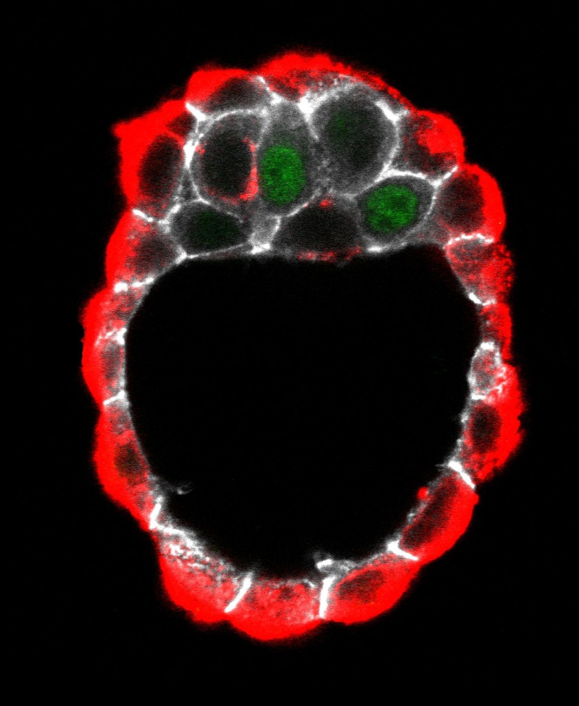
Mouse embyo
Mouse embryo at E3.75 days of development. At this stage 3 cell types are present in the embryo: the outside trophectoderm enclosing the inner cell mass that is composed of epiblast and primitive endoderm cells intermingled in a salt and pepper pattern. NANOG (green) identifies epiblast cells while LRP2 labels the outside trophectoderm as well a subset of primitive endoderm cells in the inner cell mass. E-cadherin staining is shown in white..
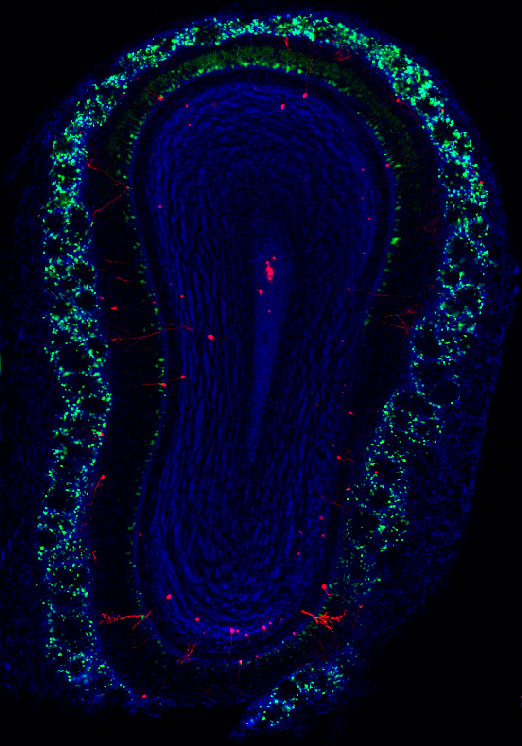
Postnatal Neurogenesis
Cross section through a mouse olfactory bulb. This brain region is one of the few where new neurons are produced at postnatal stages. Here, newborn neurons have been labeled in red by electroporation of a red plasmid within the subventricular zone. A nuclear counsterstain (blue) allows visualizing the olfactory bulb cytoarchitecture, whereas a calretinin staining (green) highlight a population of interneurons within its periphery.
